
A Machine Learning Approach for Detecting COVID-19 and Pneumonia
Introduction: The Growing Role of Machine Learning in Healthcare Diagnostics
The healthcare industry is experiencing a digital transformation, driven by advancements in machine learning (ML) and signal processing. These technologies are increasingly becoming critical in improving diagnostic accuracy, particularly for diseases such as COVID-19 and pneumonia. With the outbreak of the COVID-19 pandemic, early and accurate detection of the virus has become more important than ever. Chest X-rays are one of the primary tools used to detect lung conditions, but manually interpreting these images can be slow and subject to human error. This is where machine learning models—combined with signal processing techniques—are making significant strides.
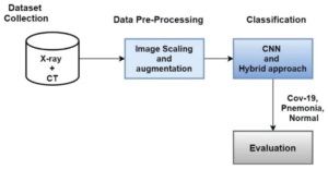
Figure: 1 Proposed Methodology Diagram.
This blog post delves into how signal processing and machine learning algorithms, particularly Convolutional Neural Networks (CNNs) and hybrid models, are being used to detect COVID-19 and pneumonia from chest X-ray images. By leveraging the power of these advanced technologies, healthcare providers can quickly and accurately diagnose these diseases, ensuring better treatment outcomes for patients.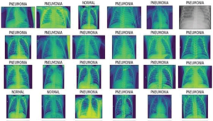
Figure: 2 Pneumonia images.
Understanding Machine Learning and Signal Processing in Medical Imaging
Before exploring how machine learning and signal processing contribute to diagnosing COVID-19 and pneumonia, it’s important to understand what these terms mean and how they’re applied in medical imaging.
What is Signal Processing?
Signal processing refers to the manipulation and enhancement of signals, such as images, to make them easier to analyze. In the case of medical imaging, signal processing techniques are used to enhance the quality of images, remove noise, and highlight key features that are important for diagnosis. For example, X-ray images of the chest may contain background noise or blur that makes it difficult for a radiologist or an algorithm to identify signs of COVID-19 or pneumonia.
Common signal processing techniques used in medical image analysis include:
- Noise Reduction: Removing irrelevant noise from images, making it easier to focus on the features that matter.
- Edge Detection: Identifying the edges of structures within the image, such as lung borders, which can be critical in detecting diseases.
- Contrast Enhancement: Improving the contrast of images to make abnormalities more visible.
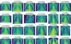
Figure: 3 Actual and predicted label images.
What is Machine Learning?
Machine learning is a subset of artificial intelligence (AI) that involves training algorithms to identify patterns in data. In medical imaging, machine learning models are trained on large datasets of labeled images (e.g., chest X-rays with annotations for pneumonia or COVID-19) to recognize patterns that indicate the presence of disease.
One of the most effective machine learning algorithms used for image recognition is Convolutional Neural Networks (CNNs). CNNs are deep learning models designed specifically for processing grid-like data, such as images. CNNs automatically learn features from raw image data, eliminating the need for manual feature extraction. This makes CNNs highly effective in medical image analysis, where subtle features in the image are critical for disease detection.
Signal Processing and Machine Learning: A Powerful Combination
While machine learning has revolutionized healthcare diagnostics, signal processing still plays a crucial role in preparing medical images for analysis. In the context of COVID-19 and pneumonia detection, the combination of these two technologies can significantly improve diagnostic accuracy.
How Signal Processing Improves Machine Learning Models
Image Enhancement:
Signal processing techniques like contrast enhancement and noise reduction help improve the quality of chest X-ray images, making it easier for machine learning models to identify important features. In cases where the abnormalities are small or subtle, enhanced images can improve the model’s ability to detect these issues.Dimensionality Reduction:
The large datasets used for training machine learning models often contain many features that are irrelevant to the task. Dimensionality reduction techniques, such as Principal Component Analysis (PCA), help reduce the complexity of the data, making it easier for the model to focus on the most important features without losing relevant information.Feature Extraction:
Signal processing techniques help extract features from images, such as lung opacity or pleural effusion, which can indicate pneumonia or COVID-19. These features are then used by machine learning algorithms to make predictions about the likelihood of disease.
Hybrid Models for Improved Detection of COVID-19 and Pneumonia
In medical image diagnostics, hybrid models combine the power of deep learning (like CNNs) with traditional machine learning algorithms. These models take advantage of both techniques to provide more robust and accurate predictions.
Hybrid Models in Action: CNNs Combined with Traditional Algorithms
CNN + Support Vector Machines (SVM):
This hybrid model combines CNNs for automatic feature extraction and SVMs for classification. SVM is a powerful classifier that works well with high-dimensional data, making it an excellent complement to CNNs. In this model, the CNN extracts features from chest X-ray images, and the SVM classifier determines whether the image shows signs of pneumonia or COVID-19.CNN + Random Forest (RF):
Random Forests are ensemble learning methods that build multiple decision trees to make classifications. When combined with CNNs, RF models provide an extra layer of classification that can improve the model’s accuracy. This hybrid model has shown to be particularly useful in pneumonia detection, where CNNs detect key image features, and RF classifies the overall image.CNN + XGBoost:
XGBoost is a highly efficient machine learning algorithm used for classification and regression tasks. When paired with CNNs, XGBoost refines the classification process by learning from the features extracted by the CNN, leading to enhanced diagnostic performance.
Performance of Hybrid Models in Pneumonia and COVID-19 Detection
The CNN model has shown remarkable results for detecting pneumonia from chest X-rays. It has achieved a 99.47% recall for pneumonia, meaning that the model is highly effective at identifying true cases of pneumonia. The hybrid CNN + RF model achieved 90.3% accuracy for pneumonia detection, showing that traditional machine learning algorithms can enhance the performance of deep learning models.
For COVID-19 detection, the CNN model achieved 95.45% accuracy, showing its strong potential for distinguishing COVID-19 from other diseases. The hybrid models, including CNN + SVM and CNN + XGBoost, also performed well but did not outperform the CNN alone.
These results highlight the effectiveness of machine learning models in detecting COVID-19 and pneumonia, particularly in high-pressure environments where quick and accurate diagnostics are crucial.
Challenges and Future Directions
While machine learning and signal processing have made significant strides in medical diagnostics, there are still several challenges to overcome:
Dataset Limitations:
Most datasets used for training models are relatively small and may not represent the diversity of global patient populations. Expanding these datasets to include more diverse patient groups is crucial for improving the generalizability of the models.Overfitting:
Overfitting occurs when a model becomes too specialized in the training data, which can lead to poor performance on new, unseen data. To prevent overfitting, techniques like regularization and cross-validation should be used during model training.Model Interpretability:
Deep learning models, especially CNNs, are often considered black-box models, meaning it is difficult to understand how they arrive at a decision. Improving model interpretability will be essential for gaining the trust of clinicians and ensuring the widespread adoption of these technologies in healthcare.Real-Time Implementation:
For these models to be used in real-time diagnostic settings, further optimization is required. Reducing computation time for inference and integrating the models into clinical workflows will be key to their success.
Conclusion: Revolutionizing Medical Diagnostics
The integration of machine learning and signal processing is transforming the way we approach COVID-19 and pneumonia diagnosis. By combining deep learning models like CNNs with traditional machine learning algorithms, we can significantly improve diagnostic accuracy and speed. This has the potential to save lives by providing timely and accurate diagnoses, especially in resource-limited settings.
As machine learning continues to evolve, its applications in healthcare will only expand, bringing us closer to a future where AI-powered diagnostics are the norm. However, addressing the challenges of data diversity, model interpretability, and real-time implementation will be critical to ensuring the widespread adoption of these technologies in clinical practice.