Fibrin Enhances an In Vitro Wound Healing Model Utilizing Fibroblast-Populated Collagen Lattices
Enhancing In Vitro Wound Healing with Fibrin in Collagen Lattices
The study of wound healing has long benefited from in vitro models that replicate the dynamic processes of tissue repair. In this post, we explore how fibrin, a vital protein in clot formation, contributes to the efficacy of wound closure in fibroblast-populated collagen lattices (FPCLs). Using these models, researchers have revealed important findings that can inform future medical treatments and wound care practices.
Key Insights from the Study
- Fibrin’s Role in Wound Healing: The research highlights how fibrin helps in accelerating wound closure when integrated into relaxed FPCLs. The presence of fibrin stabilizes the matrix and facilitates contraction, mimicking the natural wound repair environment.
- Mechanics of FPCLs: Two main models were analyzed: relaxed FPCLs and stressed FPCLs. While relaxed lattices allowed for quicker wound closure when treated with fibrin, stressed FPCLs maintained higher tension, inhibiting complete closure even with fibrin.
- Histological Observations: Under the microscope, fibrin-treated lattices showed changes in collagen organization and fiber packing that supported wound healing. The use of picrosirius red staining illustrated these structural modifications.
Methodology and Findings
The research employed hTERT-BJ1 fibroblasts cultured in collagen matrices and subjected to incisional wounds. Fibrin treatment demonstrated its potential in reducing wound size and improving repair over time, particularly in relaxed FPCLs. However, in stressed matrices, the high tension reduced the closure effect, showing the limitations of mechanical stress on fibrin efficacy.
Implications for Future Research
These findings reinforce the importance of incorporating fibrin in wound healing models to better understand tissue repair mechanisms. The study also underscores the limitations of high-tension environments in wound closure, providing a foundation for developing enhanced therapeutic strategies.
Tags
- Wound Healing
- Fibrin in Tissue Repair
- Collagen Lattices
- In Vitro Models
- Fibroblast Research
- Biomechanics of Wounds

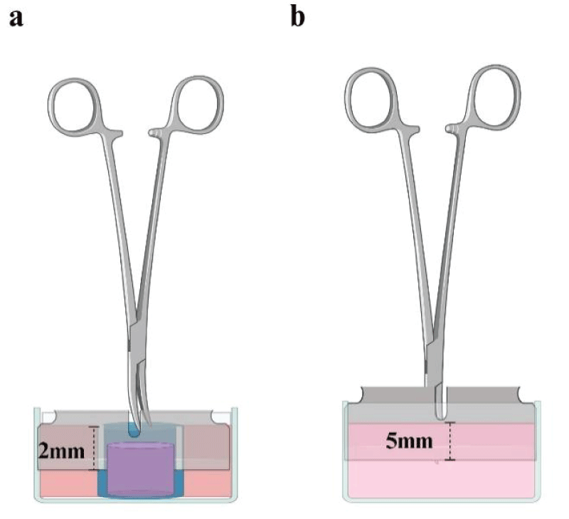
Devices to create wounds on FPCLs. Standardization of wounds was achieved through a sterilized stainless-steel knife coupled with Kelly forceps. For relaxed FPCL it was necessary to hold the lattice by introducing it into a plastic straw with a slit that would allow the knife to fit exactly 2 mm (a). For stressed FPCLs, the fit distance of the knife was 5 mm measured from the tip of Kelly forceps to the knife edge, in order to reach the exact depth into the de lattice when the tip of the forceps had touched the surface of the FPCLs (b). Created with BioRender.com.

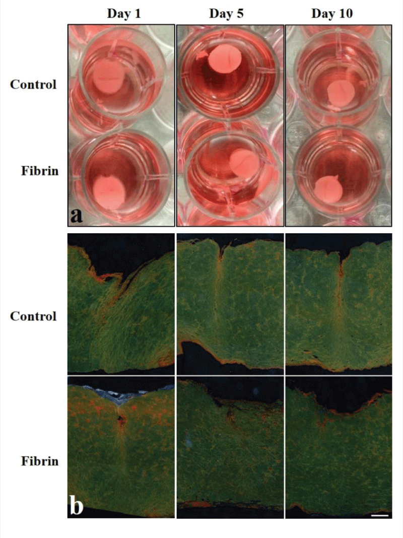
Relaxed FPCLs. a) Pictures of representative relaxed FPCL of control and fibrin treated at different time points, 1, 5, and 10 days. b) Photomicrographs of cross sections of relaxed FPCLs stained with picrosirius red. The bar represents 200 μm.